
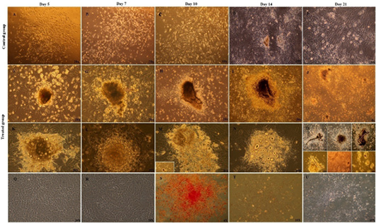
Morphology of MSCs control group subcultured in complete expansion medium and other treated subgroup sub-cultured in osteogenic medium supplemented with b-FGF during three weeks after subculture: (A-E) control group, thin and fiberoblast-like MSCs were arranged in bundles and sheets after subculture at d (A) 5, (B) 7, (C) 10 and (D, E) network pattern of growth was seen. Networks were connected to each other’s via long cellular processes at day 14 and 21(F-R) Nodule formation was observed at day 5 in osteogenic medium, and increased in size and number progressively over time during three weeks of subculture. (Q, R) Morphology of MSCs completely different from morphology of osteoblast cells, black arrow,(S) Alkaline phosphatase histochemical staining of control group; the cells were negative for ALP staining (red) at d 7,14, and 21,(T,U) control group was negative for both Alizarin red and Von Kossa staining through three week of differentiation.